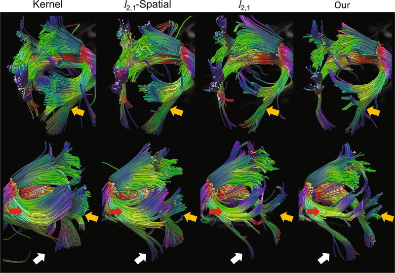
Fig. 4.
Fiber tracts of the splenium of corpus callosum, extracted from diffusion-weighted atlases at 6 months of age. The proposed atlas provides more clearly separated branches and well-connected tracts to the cerebral cortex. Kernel: kernel regression using age; l2,1-Spatial: muti-task LASSO with spatial consistency; l2,1: multi-task LASSO with spati-temporal consistency; Our: the proposed method. 1st row: right side of the splenium; 2nd row: left side of the splenium.

