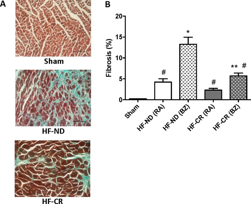
Figure 5. Long-term caloric restriction reduces left ventricular fibrosis in ischemic HF.

Masson-Trichrome staining denoting cardiac fibrosis. (A) Representative images of Sham as well as HF-ND and HF-CR in border zone (BZ). Black scale bar corresponding to 100 µm (B) Quantification of the % fibrosis in Sham as well as remote area (RA) and BZ of HF groups. N= 3 to 5 per group. Data are presented as mean±SEM. *p<0.0001 vs Sham, **p<0.05 vs Sham, #p<0.0001 vs HF-ND (BZ). One-way ANOVA analysis and Bonferroni test were used among all groups.
