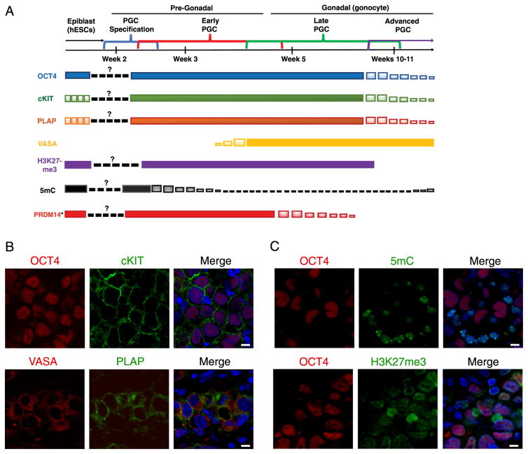
Fig. 1. Intracranial germinomas resemble late PGCs.
A. Timeline of germ cell development with expression patterns of germ line markers and epigenetic markers. *Early PGC expression pattern extrapolated from non-human primate data (Sasaki et al., 2016).
B. Immunofluorescence staining of intracranial germinomas for germ line markers, top panel: OCT4/cKIT/DAPI(merge), bottom panel: VASA/PLAP/DAPI(merge). Scale bars, 8 μm.
C. Immunofluorescence staining of intracranial germinomas for epigenetic markers, top panel: OCT4/5mC/DAPI (merge), bottom panel: OCT4/H3K27me3/DAPI(merge). Scale bars, 8 μm.

