
Figure 5. Duodenum histology of mice with rotavirus infection.
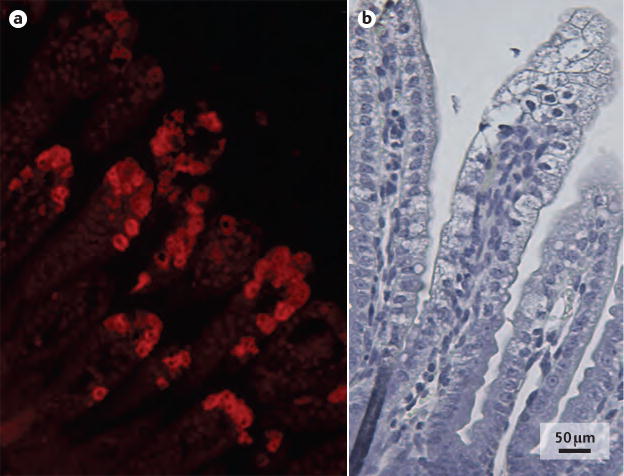
Histopathological images of the duodenum of a mouse pup infected with a murine rotavirus strain (EDIM), 48 hours after infection. a | Rotavirus predominantly infects mature enterocytes at the middle and top of intestinal villi indicated by immunofluorescent labelling of rotavirus antigen viral protein 6. b | Vacuolization of enterocytes in the top and middle of intestinal villi can be observed with rotavirus infection, but crypt cells are unaffected.