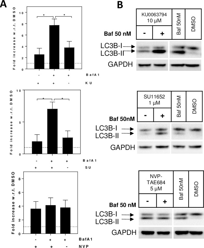
Fig 5. Co-treatment with bafilomycin A1 to distinguish autophagy enhancers versus blockers.
Rat primary astrocytes were treated with 10 μM KU0063794, 1 μM SU11652 or 5 μM NVP-TAE684 (concentration selected from information in Fig 2) followed by either 50 nM bafilomycin A1 (+Baf) or vehicle (DMSO, -Baf) for an additional 4 hours. Control samples were treated with vehicle (DMSO) for 2 hours followed by either bafilomycin A1 (50 nM) or vehicle (DMSO) for an additional 4 hours. LC3B-II TR-FRET signals are reported as fold increase with respect to the vehicle. Co-treatment of KU0063794 and bafilomycin A1 increased LC3B-II, compared to KU0063794 or bafilomycin A1 alone (N = 2, one-way ANOVA, p<0.01; Tukey’s multiple comparison test, *p<0.05); co-treatment of SU11652 and bafilomycin A1 increased LC3B-II, compared to SU11652 or bafilomycin A1 alone (N = 2, one-way ANOVA, p<0.01; Tukey’s multiple comparison test, *p<0.05); co-treatment of NVP-TAE684 and bafilomycin A1 did not alter the LC3B-II signal (N = 2, one-way ANOVA, p>0.05; (A). Western blots (B) confirm the TR-FRET data.

