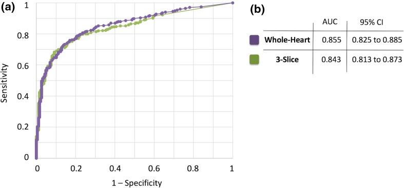
Fig. 3.
Comparison of the diagnostic accuracy of whole-heart myocardial perfusion imaging (MPI) versus 3-slice MPI with invasive coronary angiography (ICA) as the reference. a Receiver-operating characteristic (ROC) curves corresponding to whole-heart imaging (using the whole-heart TPD scores) versus 3-slice imaging (using the 3-slice TPD scores) for detection of significant CAD, defined as ≥ 70% coronary stenosis (any main epicardial vessel or branches with diameter ≥ 2 mm) based on ICA (n = 651). b The area under the ROC curves (AUCs) for whole-heart imaging and 3-slice imaging are 0.855 and 0.843, respectively (P = 0.07), indicating a statistically insignificant difference in terms of diagnostic performance. The 95% confidence interval corresponding to each AUC is also provided in panel (b). CAD coronary artery disease, TPD total perfusion deficit

