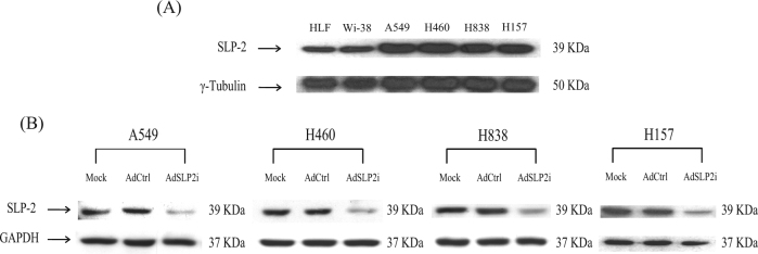
Fig. 1. The expression of SLP-2 after AdSLP2i transfection in various cell lines.
a SLP-2 expression levels in normal and NSCLC cells were analyzed by western blotting using antibodies against SLP-2 and γ-tubulin. The primary antibodies used are shown on the left of the panel. The cell lines are shown on the top of the panel. b Western blot assays for the expressions of SLP-2 protein in A549, H460, H838, and H157 NSCLC cells on day 3 after treatment with mock infection (Mock), or with either AdCtrl or AdSLP2i at m.o.i. of 100

