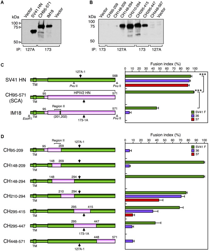
FIGURE 4.

Middle region of the SV41 HN protein is not required for activating the SV41 F-like chimeric proteins. (A,B) Detection of cell surface-localized HN proteins. Subconfluent BHK cells in six-well culture plates were transfected with 2 μg/well of the pcDL-SRα expression vector encoding each HN protein. At 24 h post transfection, the transfected cells were biotinylated, the cell lysates were subjected to immunoprecipitation (IP) with MAb 173-1A (173) or MAb 127A-1 (127A), the precipitates were subjected to SDS-PAGE under reducing conditions, and the HN protein bands were detected by ECL as described in the section “Materials and Methods.” Vector: pcDL-SRα expression vector used as the negative control. The SV41 HN protein migrates much faster compared to CH95-571 and IM18 as reported previously (Tsurudome et al., 2015), presumably reflecting the remarkable difference in the number of potential N-glycosylation sites between the HN proteins of SV41 and HPIV2 (Tsurudome et al., 1990): the SV41 HN protein (568 aa) migrates as a 67-kDa protein (Tsurudome et al., 1989) while the HPIV2 HN protein (571 aa) migrates as an 82-kDa protein (Ito et al., 1987; Tsurudome et al., 1990). (C,D) F protein specificity of the HN proteins. The average fusion index was determined at 24 h post transfection as described in the legend for Figure 2D; error bars indicate standard deviation. The representative data of more than three independent experiments are shown. The fusion indices are not normalized to the cell surface-localization levels which could not be determined due to the use two detection antibodies. The statistical significance was evaluated by one-way ANOVA as described in the section “Materials and Methods” (∗∗∗p < 0.01, n = 10). ns, not significant. Open triangles indicate the positions of restriction enzyme sites in the cDNA encoding the SV41 HN protein or IM-18. Arrows indicate the positions of epitopes for the two anti-HN monoclonal antibodies. The numbers above each cylinder denote the residue numbers of the HPIV2 HN fragments that had replaced the corresponding part of the SV41 HN protein.
