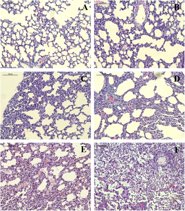
FIGURE 3.

Pathological changes of lung tissue from 6D2 infected mice. (A–F) Lung tissue from mice infected by r6D2-WT, r6D2-MA (HA), r6D2-MA (PA), r6D2-MA (PB2), r6D2-MA (PA/PB2), and r6D2-MA (HA/PA/PB2), respectively. Lung sections were subjected to hematoxylin–eosin staining and displayed at a magnification of ×200.
