
Fig. 4. Decreased expression of E2F1 protein in T cells in response to METH.
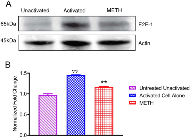
Primary human T were activated with human anti-CD3/CD28 for 48 h treated with METH (100 μM) for 24 h. a Representative immunoblots of E2F1 and internal standard actin of three independent experiments are shown in blots. b The relative fold expression of protein subunits normalized to actin are shown in the histogram. The data are presented as the mean ± SEM of three independent experiments. ∇∇P < 0.001 compared to unactivated cells;**P < 0.001 compared to activated cells