Figure 3.
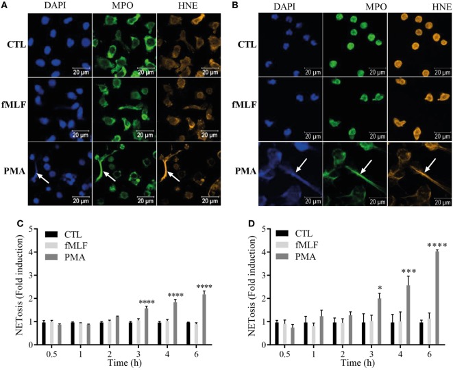
S100A8/A9 are secreted by NETosis from both differentiated HL-60 (dHL-60) cells and purified neutrophils. (A,B) Immunofluorescent staining of neutrophil extracellular traps (NETs) from dHL-60 cells (A) or neutrophils (B). dHL-60 cells or neutrophils were stimulated with 100 nM fMLF (middle panels) or 100 nM phorbol 12-myristoyl 13-acetate (PMA) (lower panels) for 4 h and stained for DNA (DAPI; blue staining), myeloperoxidase (MPO; green staining) or neutrophil elastase (HNE; orange staining). Pictures are representative of at least three independent experiments. (C,D) Quantification of NET release from dHL60 cells (C) or neutrophils (D). Extracellular NET-DNA was quantified with the cell-impermeable fluorescent dye Sytox blue and NET-release was detected via fluorescence emission. NET formation was normalized to the non-stimulated control and expressed as fold induction. Results are presented as mean ± SEM of four independent experiments. *p < 0.05; **p < 0.01; ***p < 0.001; ****p < 0.0001.

