Figure 1.
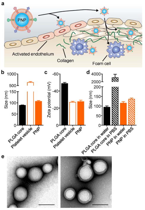
Platelet membrane-coated nanoparticle (PNP) schematic and characterization. (a) PNPs express a variety of surface markers capable of targeting different components of atherosclerotic plaques, including activated endothelium, foam cells, and collagen. (b) Z-average size of bare PLGA cores, platelet membrane-derived vesicles, and PNPs as measured by dynamic light scattering (DLS) (n = 3, mean ± SD). (c) Surface zeta potential of bare PLGA cores, platelet membrane-derived vesicles, and PNPs as measured by DLS (n = 3, mean ± SD). (d) Z-average size of bare PLGA cores and PNPs in water or in PBS (n = 3, mean ± SD). (e) Transmission electron microscopy (TEM) image of bare PLGA cores (left) and PNPs (right) negatively stained with uranyl acetate (scale bars = 100 nm).

