Fig. 1.
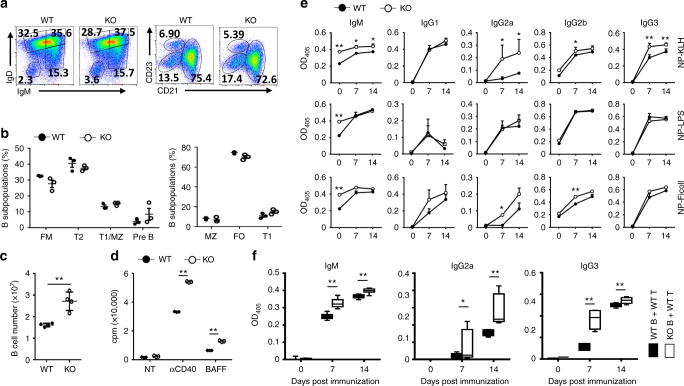
Peli1 deficiency promotes B cell proliferation and antibody secretion. a, b Flow cytometric analysis of the percentages of B cell subpopulations in the spleens of WT and Peli1-dificient mice. Data are presented as a representative plot (a) and summary graph (b). c The absolute numbers of B220+ B cells in the spleens of WT and KO mice. d Proliferation of WT and KO splenic B cells incubated in vitro for 72 h in the absence (NT) or presence of anti-CD40 (αCD40) or BAFF, then assessed by [3H]thymidine incorporation. e Enzyme-linked immunosorbent assay (ELISA) of NP-specific antibody isotypes in the serum of WT and Peli1-dificient mice immunized intraperitoneally with NP-KLH, NP-LPS, or NP-Ficoll. f ELISA of NP-specific IgM, IgG2a and IgG3 in the serum of Rag1-dificient mice that transferred with WT T cells plus WT or KO B cells, and then immunized intraperitoneally with NP-KLH. Data are shown as the mean ± SEM based on three independent experiments. Two-tailed Student’s t-tests were performed. *P < 0.05 and **P < 0.01

