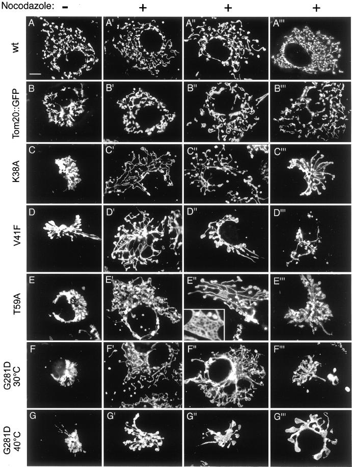
Figure 2.
Range of mitochondrial distribution defects induced by different mutations in Drp1. The left column of images shows transfected cells that were not treated with nocodazole. The remaining three columns show different examples of transfected cells that were treated with nocodazole. (A–A‴) COS-7 cells transfected with a wild-type Drp1 construct. (B–B‴) Cells transfected with a Tom20::GFP construct, which was used as a negative a control, because it causes mitochondrial clustering without affecting connectivity. (C–C‴) Cells transfected with a Drp1(K38A) construct. (D–D‴) Cells transfected with a Drp1(V41F) construct. The cell shown in D′ was grown at 30°C, whereas the cells shown in D" and D‴ were grown at 40°C. (E–E‴) Cells transfected with a Drp1(T59A) construct. The inset in E" shows an enlargement of a net formed by the mitochondria in this cell. (F–F‴) Cells transfected with a Drp1(G281D) construct and grown at 30°C. (G–G‴) Cells transfected with a Drp1(G281D) construct and grown at 40°C.

