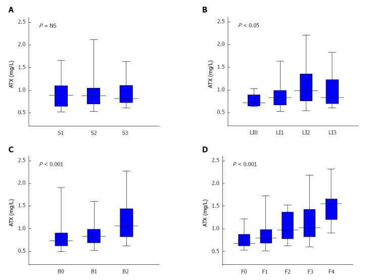
Figure 2.

Relationship between autotaxin and histological grade in non-alcoholic fatty liver disease patients for steatosis (A), lobular inflammation (B), ballooning (C), and fibrosis (D). Table 1 presents the number of subjects for each histological stage. The Kruskal-Wallis test was used for multi-group simultaneous comparisons. P values are displayed in the upper left of each graph. ATX: Autotaxin; NAFLD: Non-alcoholic fatty liver disease; NS: Not significant.
