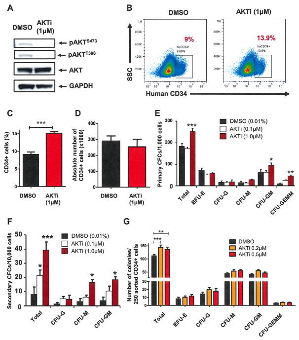
Figure 2.
Pharmacological inhibition of AKT maintains human HSPCs and enhances their colony-forming capability. (A) Inhibiting AKT activity in CD34+ cells using an AKTi decreased AKT phosphorylation at both serine 473 and threonine 308. Western blot analysis of AKT phosphorylation in CD34+ cells 24 hours after DMSO or AKTi (1 μmol/L) treatment is shown. (B) Flow cytometric analysis of human CD34 expression 7 days after AKTi (1 μmol/L) or DMSO treatment. (C) Inhibition of AKT maintains the immature surface marker of human HSPCs in vitro. The percentage of human CD34+ cells in expanding medium supplemented with vehicle (DMSO) or AKTi (1 μmol/L) is shown (***p < 0.001, n = 3). (D) Absolute number of human CD34+ cells in expanding medium was measured 7 days after AKTi (1 μmol/L) or DMSO treatment (p > 0.05, n = 3). (E) Inhibition of AKT activity enhances colony formation of human CD34+ cells. CFC assays were performed using equal numbers of cultured cells treated with DMSO (0.01%) or AKTi (0.1 and 1 μmol/L) for 7 days (*p < 0.05, **p < 0.01, ***p < 0.001, n = 3). (F) The 10,000 cells collected from primary colonies used in the secondary CFC assays (*p < 0.05, ***p < 0.001, n = 3). (G) CFC assays performed using 250 sorted CD34+ cells 7 days after DMSO (0.01%) or AKTi (0.2 and 0.5 μmol/L) treatment (**p < 0.01, ***p < 0.001, n = 3).

