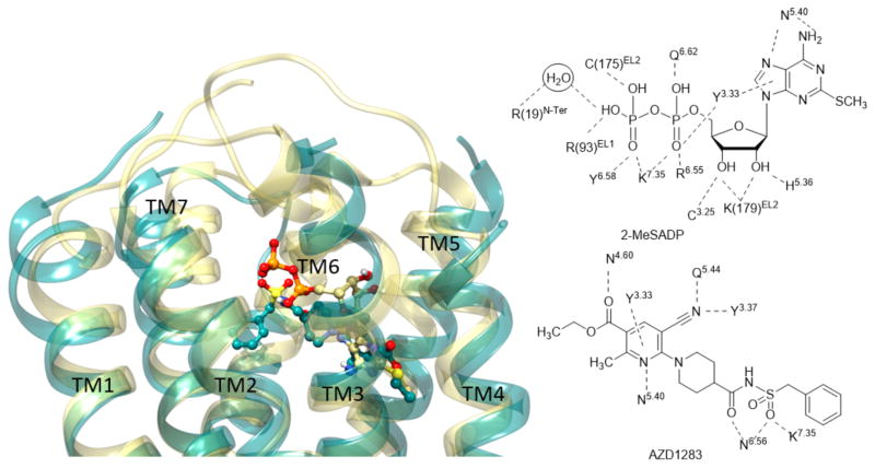
Figure 5.
Left panel: Superimposition between X-ray crystallographic structures of the hP2Y12R in complex with 2-MeSADP (28) (yellow ribbon representation for the receptor and yellow carbon atoms for the ligand) [21] and AZD1283 (35) (dark cyan ribbon representation for the receptor and dark cyan carbon atoms for the ligand) [20]. Right panel: Contacts between two co-crystallized ligands and the hP2Y12R.

