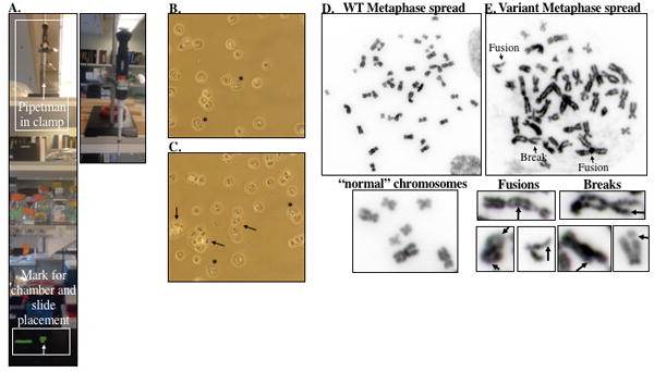
Figure 4.

A. Picture of the pipetman clamp setup for dropping cells for metaphase preparation and micronucleus analysis. The pipetman is held in a clamp that is placed on a shelf about 3 ft above the bench surface. A mark is placed on the bench top for appropriate placement of the slides. B. Bright field images using a 20X objective of metaphase spreads prior to mounting. Cells in metaphase are indicated by (*). The arrows point to clumps of cells stuck together. Checking the slides after dropping cells serves both to adjust the volume if the cells are too sparse or confluent and to get an idea of how many cells are in metaphase prior to mounting. D-E. Representative image of metaphase spreads from a cell expressing WT or BER variant enzyme taken using a 100X objective on an Olympus BX50 research microscope with QImaging Retiga 2000R digital camera. The WT metaphase spread (D) contains “normal” chromosomes without aberrations. However, the BER variant metaphase spread (E) contains multiple chromosomes with aberrations, including breaks and fusions, as indicated in the image. Additional representative chromosomes with fusions or breaks are included below (E) to serve as a guide for scoring aberrations.
