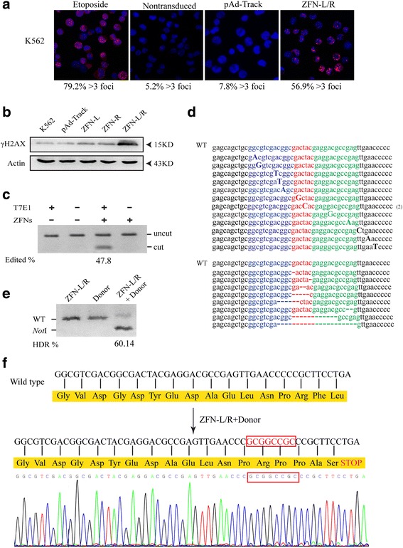
Fig. 2.

ZFNs induced gene editing of bcr-abl gene. a K562 cells were treated with 1 μM etoposide (positive control), pAd-Track or ZFN-L/R for 48 h and ZFNs-induced DSBs were detected by 53BP1 immunostaining. Nontransduced cells as negative control. The rate of cells containing more than 3 foci was shown beneath each panel. b K562 cells were transfected with pAd-Track, ZFN-L, ZFN-R plasmids separately or together of ZFN-L and ZFN-R (ZFN-L/R). The amount of γH2AX in each group was quantified by western blot. The arrows indicate the marker proteins. c ZFN-mediated gene editing revealed by T7E1 assay and the results indicated by agarose gel eletrophoresis. The bcr-abl was subjected to digestion with T7E1 to confirm the exist of insertions/deletions. Gene modification was only detected in cells transfected with ZFNs shown as ‘cut’ bands. d The genomic ZFNs target site in K562 cells was sequenced. The result showed the ZFN-induced insertions and deletions around the target region of bcr-abl. e The bcr-abl gene editing efficiency was quantified by NotI restriction enzyme. The genomic DNA of cells transfected with ZFN-L/R, Donor individually or together was extracted and amplified by PCR, then treated with NotI restriction enzyme. “WT” indicates the position of wild type PCR product and “NotI” indicates the position of the fragments generated by NotI digestion. Numbers below the lanes with NotI fragment indicate the rate of PCR product modification. f In silico analysis of sequence of ZFN-L/R and donor treated cells. The result showed the 8-bp (GCGGCCGC) insertion lead to a stop codon and a termination of translation
