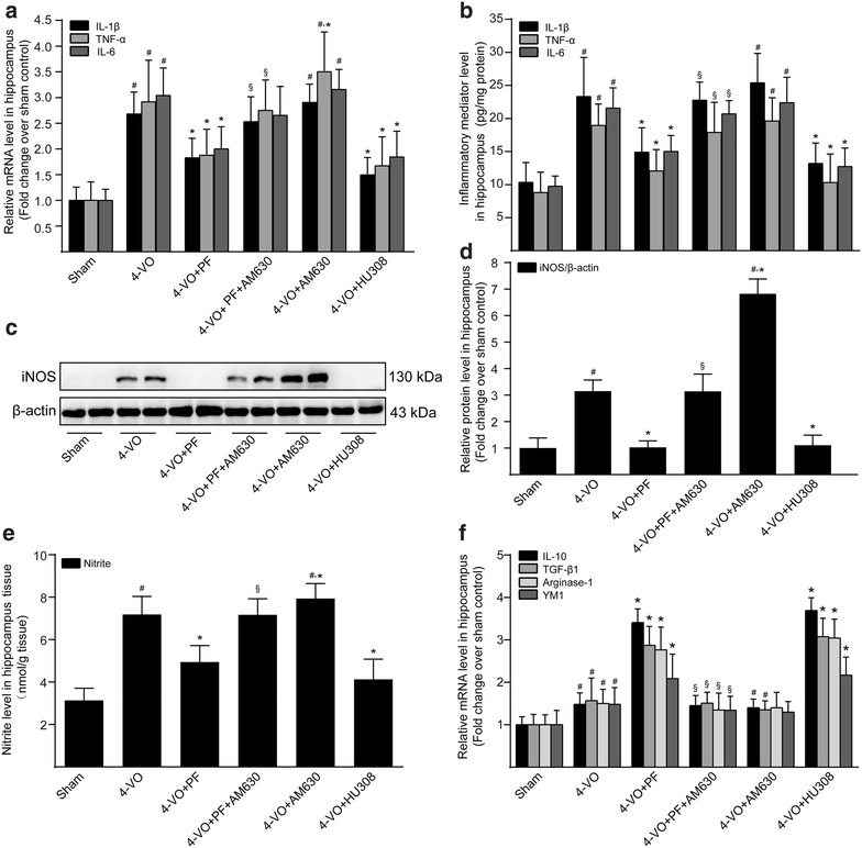
Fig. 3.

Effects of paeoniflorin on expression products of microglia/macrophage in hippocampi of rats after cerebral ischemia. One week after four-vessel occlusion (4-VO) surgery, rats were intraperitoneally administered saline (4-VO), paeoniflorin (4-VO+PF; 40 mg/kg/day), paeoniflorin+AM630 (4-VO+PF+AM630; 40 + 3 mg/kg/day), AM630 (4-VO+AM630; 3 mg/kg/day) or HU308 (4-VO+HU308; 3 mg/kg/day) for consecutive 28 days. a After treatment, real-time quantitative PCR analysis and b ELISA were used to detect the mRNA and protein levels of the proinflammatory cytokines IL-1β, TNF-α and IL-6, respectively. c Western blotting analysis was performed to further detect iNOS protein expression level. β-Actin served as an internal control. d The protein level of iNOS was expressed as arbitrary densitometric unit and normalized by the value of β-actin and finally expressed relative to the protein level in the sham-operated group (defined as 1-fold). e The content of nitrite in the hippocampus tissues of rats were determined by the Griess reaction. f Real-time quantitative PCR analysis was used to detect the mRNA levels of the anti-inflammatory cytokines IL-10 and TGF-β1; and the M2-associated markers arginase-1 and Ym1. Each bar represents mean ± SD of three independent experiments. n = 6 rats per group. #P < 0.05 versus sham group, *P < 0.05 versus 4-VO model group, §P < 0.05 versus 4-VO+PF group
