Fig. 1.

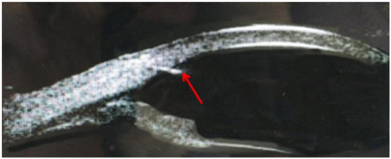
Anterior segment optical coherence tomography (OCT) image of the affected cornea. The red arrow is pointing at the foreign body in the temporal limbus of the left eye

Anterior segment optical coherence tomography (OCT) image of the affected cornea. The red arrow is pointing at the foreign body in the temporal limbus of the left eye