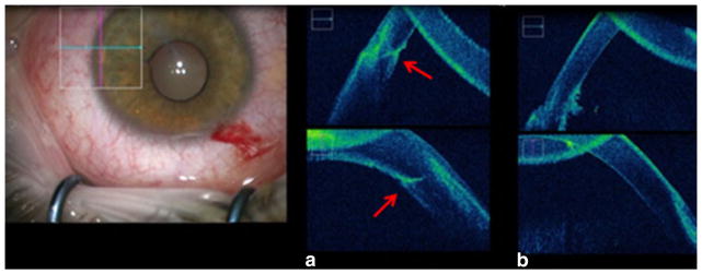
Fig. 2.
Intraoperative photograph from the operating microscope (surgeon’s view). The OCT images (a, b) correspond to the cornea and limbus focused in the rectangular box shown. a Intraoperative OCT image of the affected cornea before the surgical removal of the foreign body. Horizontal cross-sectional image (top) and vertical cross-sectional image (bottom) with the red arrows pointing at the foreign body. b Intraoperative OCT image of the affected cornea confirming the complete removal of the foreign body from the temporal limbus. Horizontal cross-sectional image (top) and vertical cross-sectional image (bottom)

