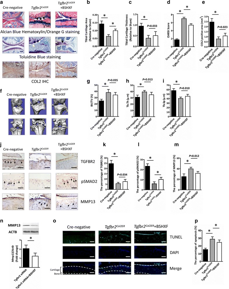
Fig. 4.

BSHXF decelerates cartilage degeneration in Tgfbr2Col2ER mice via down-regulations of MMP13. a Histological knee joint sections (100×) stained using Alcian Blue Hematoxylin/Orange G and Toluidine Blue, and COL2 IHC. Joint degradation are labeled (black arrows: cartilage degradation, blue arrows: osteophyte formation, purple arrows: subchondral sclerosis). Cartilage architecture was evaluated using the Osteomeasure System to determine b the tibial cartilage area, c tibial cartilage thickness, d OARSI scoring and e COL2 positive area. f Representative micro-CT images. Quantification of the g BV/TV, h Tb. Th and i Tb. Sp by static histomorphometry. j Knee joint representative IHC image (200×) of TGFBR2, pSMAD2 and MMP13 stained chondrocytes (brown; black arrows) with cell nuclei counterstained with hematoxylin (blue). Quantification of k TGFBR2, l pSMAD2 and m MMP13 as positive cells rate. n Mmp13 mRNA and protein expression inchondrogenic ATDC5 cells. o Terminal deoxynucleotidyl transferase dUTP nick end labeling chondrocyte apoptosis (200×). p Quantification of TUNEL staining positive cells was performed to evaluate chondrocyte apoptosis. Bars represent mean ± SD (n = 10). *P < 0.01. Scale bars = 200 μm
