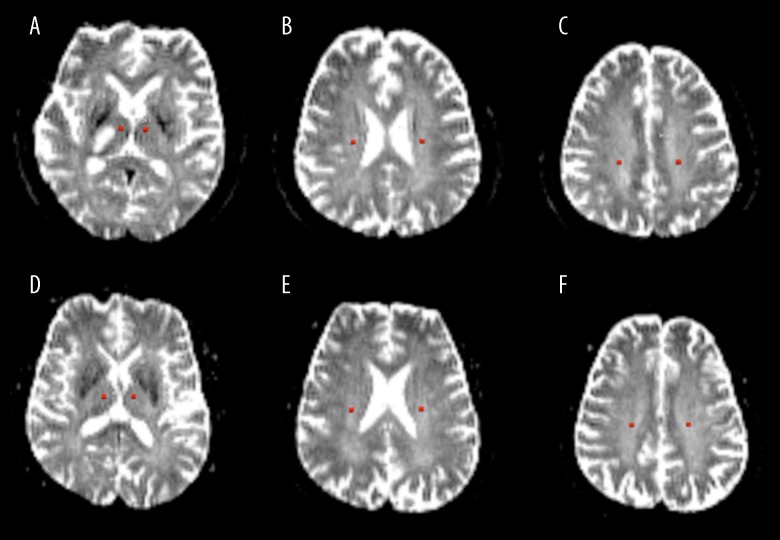
Figure 1.
Definition of regions of interest (ROIs) were defined a 4-voxel circle on ADC images (b=0). For the patients’ contralesional side (left side of images A–C) and control group (images D–F), ROIs were placed along the thalamic radiata fiber pathway in both left and right side at 3 positions: thalamus, corona radiata, and semiovale. For the patients, the ROIs in the ipsilesional side (right side of images A–C) were carefully placed to avoid the ischemic lesion and the thalamic fibers connecting to the lesion. There was no abnormal signal outside the ischemic lesion in patient images or in control subject images.

