Figure 1.
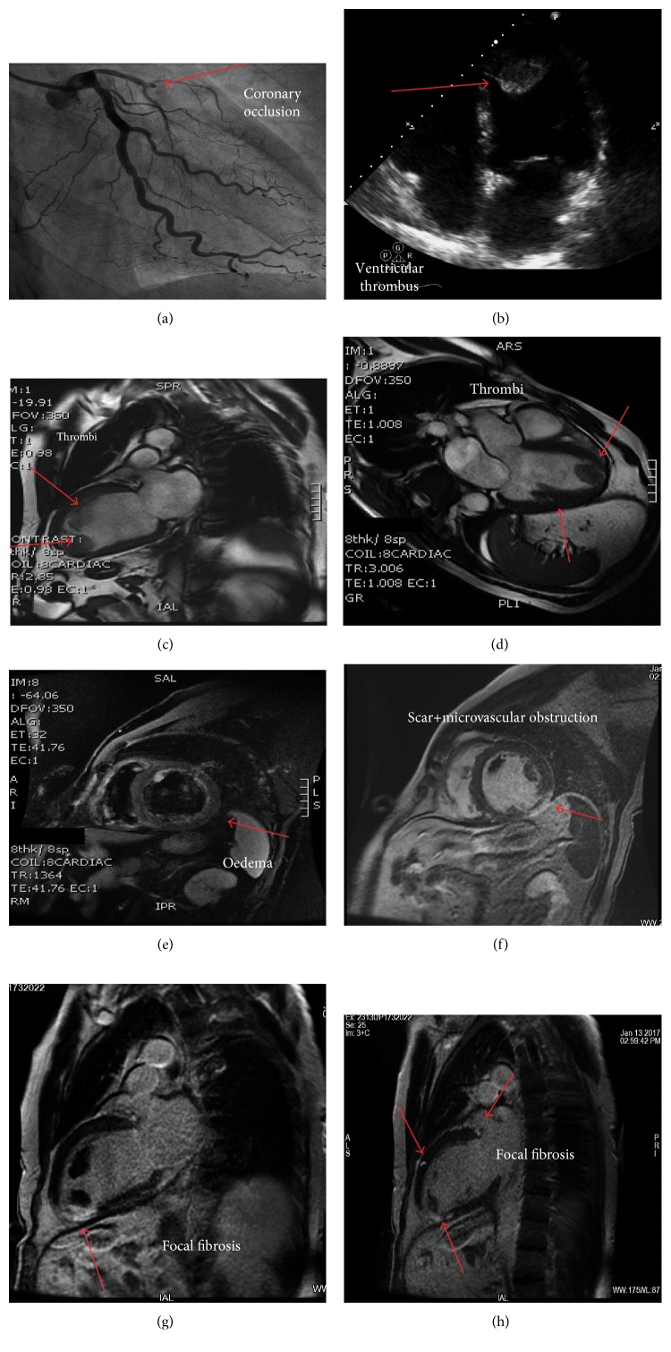
(a) Coronary angiography showing the occlusion of the left descending artery in the proximal segment (arrow); (b) transthoracic 2-D echocardiography showing an apical ventricular thrombus; (c, d) cardiac magnetic resonance (CMR) unveiling the presence of two large left ventricular thrombi in the apex and along the anterior wall; (e) CMR TIR-T2 sequences showing myocardial edema involving the anterior wall of the left ventricle; (f, g) delay enhancement revealing scar, microvascular obstruction, and fibrosis (arrows); (h) CMR imaging: three hyperenhancement focal areas of fibrosis.

