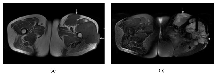
Figure 1.
A 43-year-old female with a moderately differentiated adult fibrosarcoma. (a, b) Axial SE T1WI and fat suppressed FSE T2WI, exhibiting two lobulated long T1 mixed T2 signal masses in the left hip deep fascia. The band-like areas of low signal suggest tumor fibrous septa (black arrow in (b)), and the patch-shaped regions of long T1 and T2 signal in the left gluteus maximus muscle represent muscle edema (asterisk in (b)). The lesion broke through the deep fascia (white arrows in (a), (b)).

