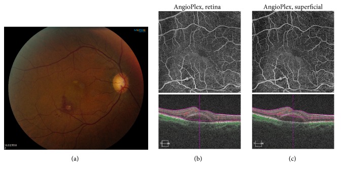
Figure 1.
A 70-year-old female presented with decreased vision in the right eye. (a) Fundus photography shows the retinal arterial macroaneurysm (RAM) along the infratemporal vascular arcade, with surrounding subretinal hemorrhage and fluid. (b) TOP: optical coherence tomography angiography (OCT-A) retina slab clearly delineates the RAM. BOTTOM: spectral domain optical coherence tomography (SD-OCT) shows submacular fluid. (c) TOP: OCT-A superficial slab reveals the RAM. Bottom: SD-OCT shows submacular fluid.

