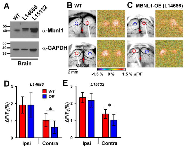
Fig. 8.
Decreased contralateral response in mice over-expressing MBNL1. A. Expression of MBNL1 in transgenic overexpression mouse lines. Western blot analysis of protein extracted from the brain of WT, L14686 (original) and L15132 (newly developed) transgenic mouse lines. Blots were probed with either an antibody against Mbnl1 (A2764, gift from Dr. Charles Thornton, University of Rochester) or GAPDH (Millipore, MAB374). B. Example images from a WT mouse where 10 Hz stimulation was performed on the right (top) and left (bottom) hemispheres. C. Images from an MBNL1-OE mouse (L14686). Responses on the contralateral hemisphere were significantly smaller regardless of which hemisphere was stimulated. D and E. Quantification of the peak flavoprotein responses from two different MBNL1-OE lines: L14686 (D, WT=8, OE=8); and L15132 (E, WT=6, OE=7). Both MBNL1-OE lines showed a significant decrease in the contralateral response compared to WT mice.

