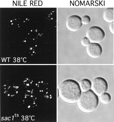
Figure 7.
sac1ts cells accumulate lipid droplets at the restrictive temperature. WT and sac1ts cells were incubated at 38°C for 2 h, fixed in 3% gluteraldehyde, and stained with Nile red (see MATERIALS AND METHODS). On the right, identical fields were observed with Nomarski optics. All cells shown are representative of >95% of cells observed.

