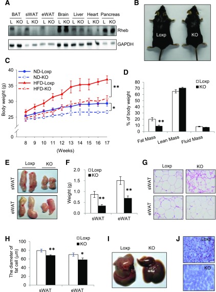
Figure 1.
RhebfKO mice are lean and resistant to HFD-induced obesity. A: Western blot analysis of Rheb expression in 6-week-old RhebfKO (KO) and Loxp (L) control mice. GAPDH was used as a loading control. eWAT, epididymal WAT. B: Representative images of mice after a 9-week HFD feeding regimen starting at 8 weeks of age. C: Body weight of mice (mean ± SEM, n = 6 – 8/group) fed the ND or HFD beginning at 8 weeks of age. D: Body composition of HFD-fed KO (n = 6) and Loxp control littermates (n = 8). E: Representative images of fat pads from HFD-fed mice. F: Weights of sWAT and eWAT of HFD-fed KO (n = 6) and Loxp control (n = 8) mice. G: Representative images of hematoxylin and eosin staining of sWAT and eWAT sections from HFD-fed KO and Loxp control mice. H: The average diameters of fat cells in KO and control mice were analyzed and quantified by the Image-Pro Plus 6.0 software. I: Representative images of liver from HFD-fed mice. J: Representative images of Oil Red O staining of liver sections from HFD-fed KO and Loxp control mice. For D–J, mice were fed the HFD for 17 weeks. Scale bars: 50 μm. Data are mean ± SEM. *P < 0.05; **P < 0.01.

