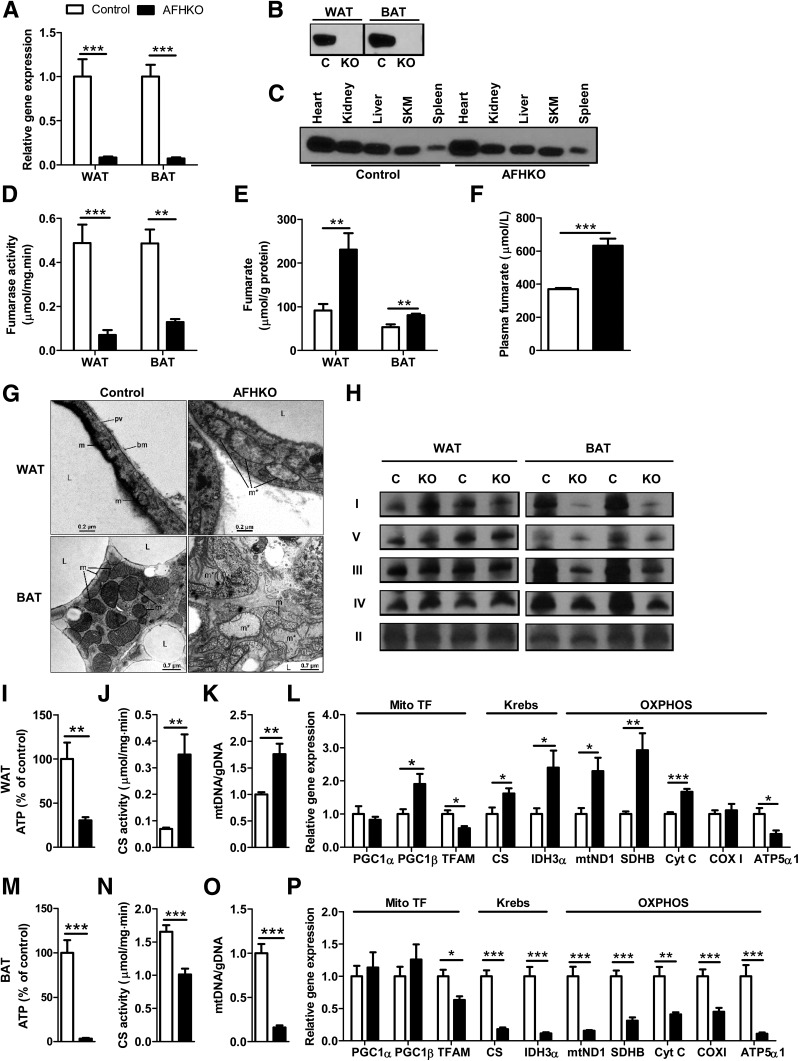
Figure 1.
AFHKO mice have adipose-specific FH gene excision. A: FH mRNA expression in perigonadal WAT and BAT assayed by real-time PCR (n = 8). B and C: Western blot for FH. Mitochondria were prepared from isolated adipocytes from perigonadal WAT and from BAT of AFHKO and control mice (10 μg mitochondrial protein/lane) (B). FH expression in nonadipose tissues (30 μg protein of tissue homogenate protein/lane) (C). D and E: FH activity and fumarate content of perigonadal WAT and BAT (n = 6). F: Plasma fumarate (5-month-old males, n = 8). G: Representative mitochondrial ultrastructure of perigonadal WAT and BAT from 5-month-old control and AFHKO mice (n = 5). H: Blue Native PAGE analysis of OXPHOS complexes. Fifteen micrograms of mitochondrial protein were used for WAT and 7.5 μg for BAT (n = 4, two representative results shown). I–L: Perigonadal WAT: ATP content (n = 6) (I), CS activity (n = 6) (J), mtDNA content (n = 8) (K), and expression levels of genes related to mitochondrial function (n = 8) (L). M–P: BAT as evaluated as in panels I–L and in samples from the same mice. *P < 0.05; **P < 0.01; ***P < 0.001. bm, basal membrane; C, control; gDNA, genomic DNA; I, complex I; II, complex II; III, complex III; IV, complex IV; KO, AFHKO; L, lipid droplet; m, mitochondrion; m*, giant mitochondrion; pv, pinocytotic vesicle; SKM, skeletal muscle; TF, transcription factor; V, complex V.

