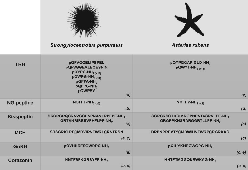
Figure 1.
Echinoderm neuropeptides that have provided new insights into the evolution of neuropeptide signalling systems. The sequences of sea urchin (S. purpuratus) and starfish (A. rubens) representatives of six selected neuropeptide types are shown. Predicted or confirmed post-translational modifications, including conversion of an N-terminal glutamine (Q) to a pyro-glutaminyl (pQ) residue and conversion of a C-terminal glycine (G) to an amide group (-NH2), are depicted and cysteine (C) residues that form or are predicted to form a disulphide bridge are underlined. Numbers in parentheses represent the number of copies of the neuropeptide in the corresponding precursor if this is greater than one. The image of S. purpuratus was obtained from https://openclipart.org/detail/170807/sea-urchin-silhouette, while the image of A. rubens was created by M. Zandawala (Stockholm University). Key: TRH: thyrotropin-releasing hormone; MCH: melanin-concentrating hormone; GnRH: gonadotropin-releasing hormone. References: (a) [54]; (b) [59]; (c) [60]; (d) [61]; (e) [62].

