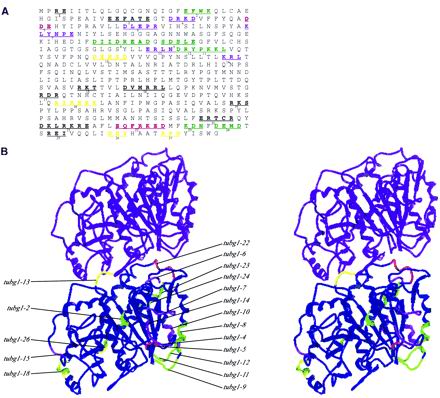
Figure 1.
Locations of the charged clusters of human γ-tubulin. (A) Amino acid sequence of human γ-tubulin is shown with the charged clusters underlined and the residues that were changed to alanine in bold letters. A cluster was defined as two or more charged residues within five residues of each other. Twenty-seven such charged clusters were altered by site-directed mutagenesis, replacing the residues with the neutral amino acid with a small side chain, alanine. The color of each cluster represents the phenotypic outcome after its expression in either wild-type S. pombe (hence a dominant phenotype), or in haploid S. pombe cells in which the endogenous γ-tubulin gene is disrupted (hence the recessive phenotype). Green, recessive lethal; purple, recessive cold sensitive; red, dominant cold sensitive; yellow, recessive lethal and dominant cold sensitive. (B) Stereo pair of the minimal free energy γ-tubulin homology model shown in longitudinal contact with the α-tubulin subunit of the microtubule minus end. The model is oriented to reveal a view from inside the lumen of a microtubule. The left and right sides thus represent putative lateral contact sites of γ-tubulin with other tubulin subunits. The colors of these clusters correspond to the color scheme showed in A.

