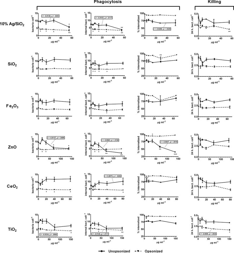
Figure 4.

NP panel F. tularensis phagocytosis and killing screen, scanning cytometry, dose response. For phagocytosis experiments, adherent PMA-matured THP-1 cells were incubated for 4 hours with indicated concentrations of ENMs prior to incubation for 2 hours with F. tularensis. For bacterial killing experiments cells were incubated for 2 hours with F. tularensis, then treated for 4 hours with ENM suspensions, washed, and incubated overnight (total 24 hours). Phagocytosis (total bacteria per cell, internal bacteria per cell, and percent internalized) and bacterial killing (bacteria per cell at 24 hours) are indicated for unopsonized (solid lines) and unopsonized (dashed lines) beads. * = p<0.05; ** = p<0.01; ***=p<0.001; Statistically significant dose/response correlation coefficients (r) and P values are indicated in inset boxes.
