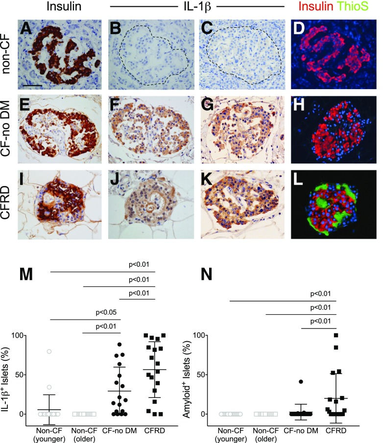
Figure 1.
IHC for insulin, IL-1β, and amyloid in subjects with and without CF. Representative staining of pancreas specimens from subjects in non-CF control (A–D), CF-no DM (E–H), and CFRD (I–L) groups, identifying islet β-cells using IHC for insulin (brown in A, E, and I and red in D, H, and L), IL-1β (brown; images shown from two subjects per group in B and C, F and G, and J and K), and amyloid (visualized by thioflavin S [ThioS] histochemistry; green in D, H, and L). Quantitation of the proportion of islets positive for IL-1β (M) and amyloid (N). Islet IL-1β positivity was increased in CF-no DM and CFRD subjects compared with age-matched non-CF control subjects, whereas islet amyloid deposition was only increased in CFRD subjects. Scale bar = 50 µm.

