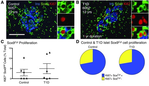
Figure 5.
Proliferative Sox9Cyt cells in adolescent and young-adult samples represent the majority of proliferating islet cells. A and B: Islet images for control (A) and T1D (B) subjects stained for insulin (Ins) (blue), Sox9 (green), and Ki67 (red). Insets indicate Ki67+ Sox9Cyt+ cells. Scale bar: 100 μm. C: Quantification of Sox9Cyt cell proliferation in a random sampling of adolescent control (n = 6) and T1D (n = 6) pancreata, represented as % total Sox9Cyt cells. Results expressed as mean ± SEM for control (n = 6) and T1D pancreata (n = 6). D: Ki67+ Sox9Cyt+ cells comprise the majority of proliferating intra-islet cells measured in a subset of highly proliferative control (n = 3) and T1D pancreata (n = 3). yr, year; yrs, years.

