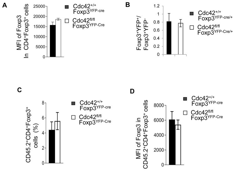
Figure 10.
Cdc42fl/flFoxp3YFP-cre mice had impaired thymocyte development but no change in nTreg differentiation. (A) Expression of Foxp3 in CD4+Foxp3+ cells from the thymus of Cdc42+/+Foxp3YFP-Cre and Cdc42fl/flFoxp3YFP-Cre mice. Data are expressed as mean fluorescence intensity (MFI). (B) The ratio of Foxp3+YFP+ vs Foxp3+YFP− cells from the thymus of Cdc42+/+Foxp3YFP-Cre/+ and Cdc42fl/fl Foxp3YFP-Cre/+ mice. (C, D) Bone marrow cells from Cdc42+/+Foxp3YFP-Cre or Cdc42fl/fl Foxp3YFP-Cre mice were mixed with bone marrow cells from BoyJ mice at 1:1 ratio and transplanted into BoyJ mice. Eight weeks later, donor-derived (CD45.2+) CD4+Foxp3+ cells in the thymus of recipient mice were analyzed for percentages of CD4+Foxp3+ cells (C) and for Foxp3 expression (MFI) (D). n = 4 or 5. Error bars indicate SD.

