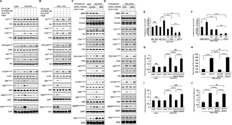
Fig. 8. ERK1/2 regulates HLA class II antibody-induced activation of intracellular signal transduction networks, cell proliferation, and migration.

EC were A, C, E, G, I infected with Ad-LacZ or Ad-CIITA or B, D, F, H, J pretreated with TNFα/IFNγ for 48 h. A, B Starved cells were pretreated with 5 μM UO126, or E, F, G, H, I, Jwith 1.0 μM UO126, or A, B, with 5.0 μM PD0325901, or E, F with 1.0 μM PD0325901, or G, H, I, J with 1.0 μM UO124 for 60 min, or C, D, Starved EC were transfected with 100 nM of MEK, or control siRNA for 48 hours. A, B, C, D Pretreated ECwere stimulated with0.1 μg/ml of HLA class II antibody for 15 min. Treatment of EC with mIgG serves as a negative control. Proteins in the pre-cleared cell lysates were separated by 6∼15% SDS-PAGE followed by immunoblotting with anti-phospho-Src Tyr418, FAK Tyr576, FAK Tyr577, p85 PI3K Tyr458, Akt Thr308, Akt Ser473, mTOR Ser2448, S6K Thr389, S6K Thr421/424, S6RP Ser240/244, or ERK Thr202/Tyr204. The membranes were re-probed with anti-Src, FAK, PI3K, Akt, mTOR, S6K, S6RP, ERK, or β-actin antibodies to confirm equal loading of proteins. E, F Cells were stimulated with 1.0 μg/ml HLA class II antibody for 48 h, incorporated with BrdU for 2 h, and harvested. EC proliferation was measured by flow cytometry. DNA synthesis S phase was gated, and proliferation index (PI) is presented as fold increase in the percentage of cells positive for BrdU normalized to negative control. G, HCells were pretreated with 10 μg/ml of mitomycin C for 2 h to inhibit cell proliferation before being assayed for their ability to migrate. A scratch wound was created with a sterile 200-μl pipette tip. Wounded cells were stimulated with 1.0 μg/ml of anti-HLA class II antibody for 16 h. The cell number between two initiated front edges was counted; migration rate was analyzed by calculating the cell number between two initiated front edges of HLA class II antibody-stimulated EC divided by the cell number between two initiated front edges of negative control EC. I, J Cell migration was measured in a transwell insert system. EC infected with Ad-LacZ or Ad-CIITA or pretreated with TNFα/IFNγwere seeded to the upper chamber of insert, pretreated with UO126 or UO124 for 60 min, and stimulated with 1.0 μg/ml HLA class II antibody for 16 h at 37°C. After incubation, the cells on the upper surface of the insert membrane were removed with a cotton swab, the migrated cells on the bottom of the insert membrane were fixed with methanol and stained with crystal violet, three middle fields per insert were photographed with 10 × objective lens, and migrated cells were counted. The bar graphs show the mean ± SEM fold change of C, D. proliferation index, E, F, G, H migrated cells. *p<0.05, **p<0.01, ***p<0.001, and ****p<0.0001 were analyzed by one way ANOVA with Fisher's LSD. Data represent at least three independent experiments.
