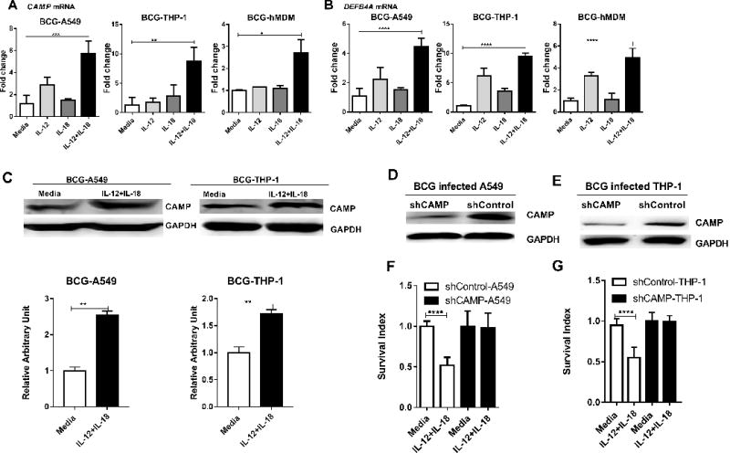
FIGURE 3. CAMP appeared to be required for the IL-12+IL-18-mediated growth inhibition of intracellular mycobacteria.

(A-B) shows mean fold changes in expression levels for CAMP(A), DEFB4A (B) in BCG-infected A549, THP-1 cells and hMDM, respectively, in the presence of media, IL-12, IL-18 or IL-12+IL-18 for 24 hours. Data are derived from 4 independent experiments using A549 or THP-1 cells, and 3 independent experiments using hMDM from 12 healthy uninfected donors. P values are calculated through ANOVA then Dunnett’s test compared with control condition: treated with culture media, * p<0.05, ** p<0.01, *** p < 0.001, **** p < 0.0001. (C) IL-12+IL-18 co-signaling led to enhanced expression of CAMP at protein levels in BCG-infected A549 (Left, Lane 1-2) and THP-1 (Right, Lane 3-4) cells. The upper panel are representative western blot analysis from 4 replicates, the lower panel are quantitative analysis of CAMP protein levels by densitometry. P values are calculated through T test compared with control condition: treated with culture media, ** p< 0.01. (D-E) shows representative western blot data demonstrating the shRNA knock-down of CAMP in A549 and THP-1 cells, respectively, compared to the shRNA-Control. (F-G) Bar graph shows that the shRNA knock-down of CAMP reverses the growth restriction of intracellular mycobacteria during IL-12+IL-18 treatment of BCG-infected A549 and THP-1 cells compared to shRNA control. Data are derived from 8 replicates in 2 independent experiments. P values are calculated through T test, **** p< 0.001.
