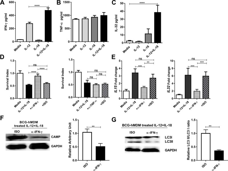
FIGURE 7. Activation of the IFN-γ-depended antimycobacterial pathway was required for the IL-12+IL-18 co-signaling-mediated growth inhibition of intracellular mycobacteria in human macrophage.

(A-C) show mean cytokine protein concentrations for IFN-γ (A), TNF-α (B) and IL-32 (C) in culture supernatants from BCG-infected hMDM in the presence of media, IL-12, IL-18 or IL-12+IL-18. Culture conditions were the same as those in Fig.1D. Cytokine proteins were measured by ELISA. Data were pooled from three independent experiments using hMDM from 12 healthy uninfected donors. P values are calculated through ANOVA then Tukey’s multiple comparisons test, **** p < 0.0001. (D) shows that IFN-γ blockade (left) but not TNF-α blockade (right) impacts mean survival indexes for BCG bacilli in IL-12+IL-18-treated hMDM. BCG-infected hMDM were treated with media or IL-12+IL-18 in the presence of neutralizing anti-IFN-γ, anti-TNF-α Ab and their isotype controls (5 ug/ml for each), respectively, and then measured for CFU counts and calculated for survival indexes as described above. In the absence of IL-12+IL-18, anti-IFN-γ or anti-TNF-α antibody does not lead to changes in intracellular mycobacterial growth (Supplementary Fig.S3A). Data are derived from 12 healthy donors in 3 independent experiments using hMDM from 12 healthy uninfected donors. P values are calculated through ANOVA then Tukey’s multiple comparisons test, **** p < 0.0001, ns, not significant. (E) shows that IFN-γ blockade-induced abrogation of intracellular BCG growth coincided with decreased expressions of IL-15 and IL-32 in BCG-infected hMDM in comparisons with the IL-12+IL-18 alone without anti-IFN-γ or isotype control. The expression levels of IL15 and IL32 mRNA in BCG infected hMDM were quantified by RT-qPCR. Data are derived from 3 independent experiments using hMDM from 12 healthy uninfected donors. P values are calculated through ANOVA then Tukey’s multiple comparisons test, *** p < 0.001, ** p < 0.01, ns, not significant. (F) The left panel: Representative western blot shows that anti-IFN-γ neutralizing Ab, not isotype control (ISO), reduced the CAMP expression during the IL-12+IL-18 treatment of BCG-infected hMDM cells; the right panel: quantitative analysis of CAMP protein levels by densitometry. Data are derived from 6 replicates. P values are calculated through T test compared with treated with isotype control, ** p<0.01. (G) The left panel: Representative western blot shows that anti-IFN-γ neutralizing Ab, not isotype control (ISO), reduced the conversion of LC3II during the IL-12+IL-18 treatment of BCG-infected hMDM; the right panel: quantitative analysis of relative LC3II/LC3I ratios by densitometry. Data are derived from 6 replicates. P values are calculated through T test compared with treated with isotype control, * p<0.05.
