Figure 4.
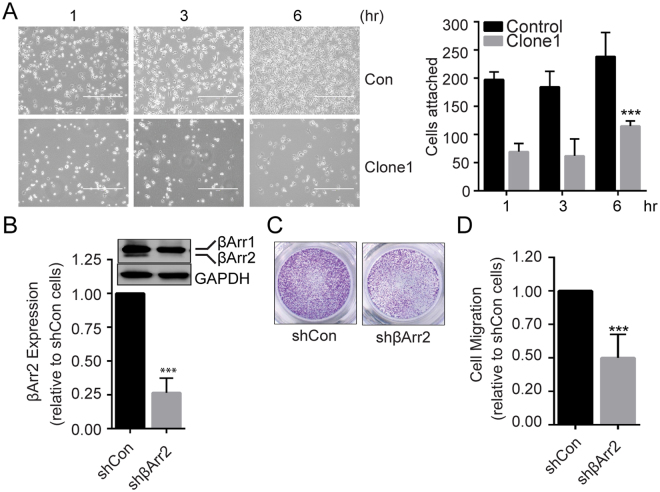
βArr2 mediates RCC cell migration and invasion. (A) Cell attachment assay. Control and βArr2ko Clone1 (2.5 × 10⁵) cells were seeded on fibronectin-coated plates, and cell attachment was observed under a phase contrast microscope at 1, 3, and 6 hr after seeding. White halos indicate semi-attached or floating cells. Bar plot showing attached cell numbers counted (10×) at each time point from three independent trials (right panel). (B) Knockdown of βArr2 in RCC cells. SN12C cells were infected with lentiviral vector containing shβArr2 or empty pLKO plasmid. Cell lysates were analyzed for βArr2 expression by Western blot (top), and band intensities were quantified by image J (bottom). (C) Cell migration assay. Control and βArr2 knockdown cells (3 × 10⁴) were starved overnight in 0.1% BSA and seeded onto transwell migration inserts. Cells that migrated through the transwell inserts over the 1% FBS gradient after 24 hr were stained with crystal violet and imaged. (D) Migrated cells in randomly-selected five fields were counted and plotted relative to control cells. Images shown are representative of three independent trials. For panels A, B and D, ***P < 0.0001.

