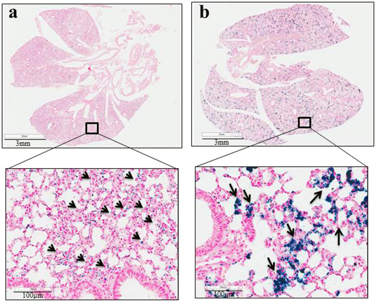
Figure 6.
Representative images of Prussian blue stained lung sections of animals treated with MNPs shows distinct patterns of iron accumulation in lungs depending upon particle coating and dose. (a) Lung tissue section from mouse injected with PEG-PEI MNPs (1 mg Fe) showing Prussian blue positive staining appears to have a dispersed pattern and concentrations of blue coloring (nanoparticle rich regions) correlate with alveolar epithelial cells, indicating nanoparticle accumulation in these cells. (b) Lung of mouse injected with CM-dextran MNPs showing a more intense clustering of particles within the interstitial spaces, a distinctly different manifestation than with PEG-PEI MNP distribution. The lower panel of figures provides a 20× magnified view of the boxed region in upper panels.

