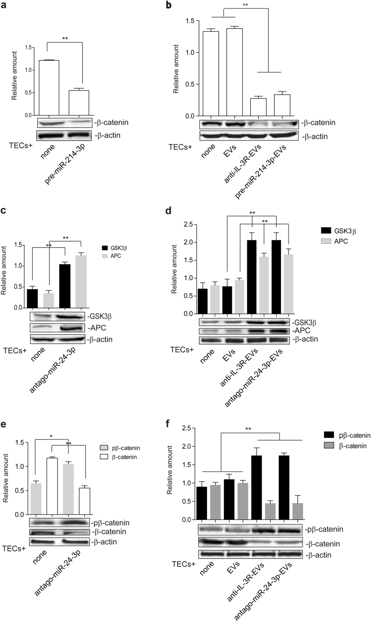
Fig. 4.
EV miR content controls β-catenin activation. a TECs that over-express miR-214-3p were subjected to SDS–PAGE to evaluate β-catenin content, normalized to β-actin. Untransfected TECs were used as internal controls (none) (n = 5) (**p < 0.01, none vs pre-miR-214-3p, unpaired t-test). b Cell extracts from TECs, left untreated or treated with EVs, anti-IL-3R-EVs or EVs enriched in miR-214-3p, were analyzed for β-catenin content, normalized to β-actin (n = 5) (**p < 0.01, none and EVs vs anti-IL-3R-EVs and pre-miR-214-3p-EVs, one-way ANOVA). c, d Cytoplasmic extracts from TECs depleted of miR-24-3p (antago-miR-24-3p) (c) and from TECs treated with antago-miR-24-3p-EVs (d) were analyzed by Western blot for APC and GSK3β content (**p < 0.01, none vs antago-miR-24-3p in (c), unpaired t-test, and **p < 0.01, EVs vs anti-IL-3R-EVs and antago-miR-24-3p-EVs in d, one-way ANOVA). e, f Cell extracts from antago-miR-24-3p TECs (e) and from TECs, treated as above (f), were analyzed for pβ-catenin and β-catenin content, normalized to β-actin (n = 4) (*p < 0.05, for pβ-catenin and **p < 0.01, for β-catenin, none vs antago-miR-24-3p in e, unpaired t-test, and **p < 0.01, none and EVs vs anti-IL-3R-EVs and antago-miR-24-3p-EVs in f, one-way ANOVA)

