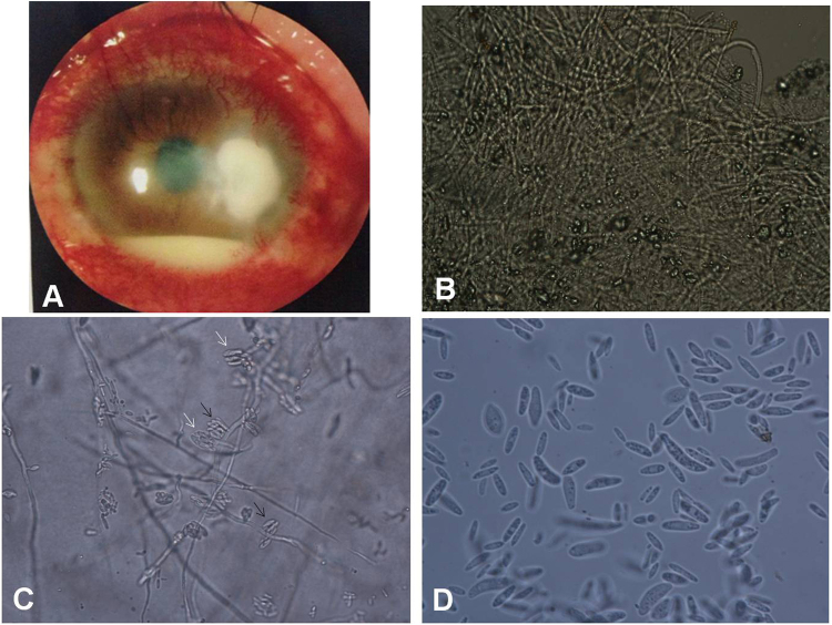
Figure 4.
The features of case 939938, which was F. proliferatum-positive. (A) Under a slit-lamp microscope, the left eye exhibited severe conjunctival injection and a next-to-central corneal ulcer (5 × 5 mm) with white, thick, irregular necrosis and irregular edges. A hypopyon of 3 mm in height was observed (×10). (B) A large number of fungal hyphae with a diameter of approximately 2 µm were observed under a microscope in corneal scrapings on 10% KOH wet film (×400). (C) A large number of false heads containing robust sickle-shaped conidia with no septa were present on both monophialides (indicated by white arrows) and polyphialides (indicated by black arrows), as shown on 10% KOH wet films of PDA after 7 d. The conidia were 4.5 × 6–15 µm in size (×400). (D) A large number of slender microconidia with no septa, sizes of 2 × 10–15 µm and some robust sickle or rod-shaped conidia (indicated by black arrows) were observed on a 10% KOH wet film of PDA after 7 d (×1000).

