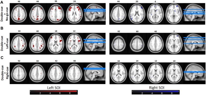
Figure 4.
Functional connectivity (psychophysiological interactions, PPIs). (A) Brain areas in which activity is more tightly coupled with activity in the source motor cortex area in the double-cue event than in the no-go-cue event. Both the left and the right Seeds of interest (SOI) showed coupling with the fronto-parietal network. The left SOI was significantly coupled with the right and left MFG, SPL, and SMA. Similarly, the right SOI was coupled with the right and left MFG, left PMd, and SPL. (B,C) Similar as 4A for the contrasts double-cue > left-cue and double-cue > right-cue, respectively. As revealed in the figures, double-cue trials have a stronger coupling with the fronto-parietal network than left/right-cue trials. Note however that the coupling is decreased for the double-cue > right-cue trials. Functionally connected areas are displayed at punc < 0.0001. Color bars represent t-statistic levels.

