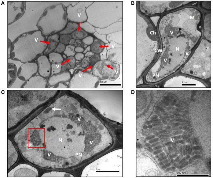
Figure 2.
Electron micrographs of thin sections of wheat leaf tissue infected with WYSV at 15 dpi. (A) Infected cells were located mainly in the vascular bundle sheath cells of leaves. (B,C) Cluster of virus particles accumulated at the perinuclear regions. A few viral particles aggregated in vesicles (white arrow) near the cell wall (panel C). (D) Magnified virus particles from section (indicate with a red box) of panel C. Ch, chloroplast; CW, cell wall; N, nucleus; PN, perinuclear regions; M, mitochondria; V, virions.

