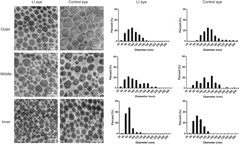
Figure 5. Collagen fibril diameter taken from representative electron micrographs (×42 000) of the LI and control eyes.
The results showed sections taken from the outer, middle and inner layers of scleral stroma and the corresponding distribution of the diameter of collagen fibrils (shown as percentage). Small fibrils have diameters ranging from 10 to 100 nm, whereas large ones ranging from 110 to 380 nm. LI: Lens-induced.

