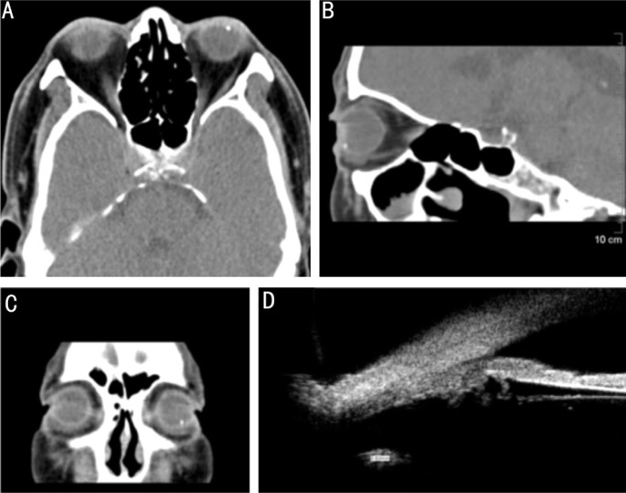
Figure 2. Confirmation of the IOFB location before surgery (patient No.4).
A-C: Coronal and sagittal position of CT-scan, the IOFB located at pars plana of ciliary; D: Ultrasound biomicroscopy (UBM)-scan showed a 0.82 mm foreign body at pars plana of ciliary, with no adhesion to surrounding tissues.

