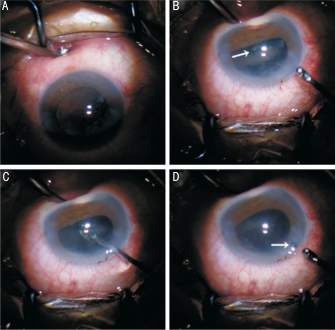
Figure 3. Scleral indentation technique applied in the IOFB removal (patient No.4).

A: Outside force to lift up the sclera around the IOFB with a chalazion curette; B: The IOFB and ciliary body in direct visualization using scleral indentation technique (white arrow, foreign body); C: Reaching the foreign body directly through the primary cornea incision and the anterior and posterior capsular opening with an IOFB forceps; D: The IOFB falling in the cornea incision when removing (white arrow, foreign body).
