Figure 7.
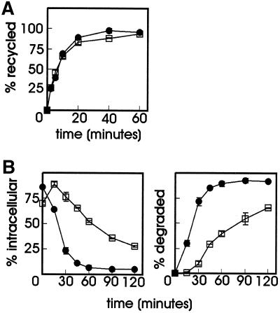
Effect of MG-132 on transferrin recycling and degradation of 125I-RAP. (A) ts20 cells were serum depleted for 1 h with or without 20 μM MG-132 and then loaded with 2 μg/ml 125I-Tf for 30 min at 30°C. Cells were chilled on ice, plasma membrane-bound 125I-Tf was removed, and the cells were incubated at 30°C in medium containing 50 μM desferal in the absence or presence of MG-132. The release of 125I-Tf was determined and expressed as a percentage of the total amount of radioactivity loaded in the cells. ●, control; □, MG-132. (B) LRP-null CHO cells stably transfected with mLRP4T100 were incubated for 1 h at 37°C with or without 20 μM MG-132 before 5 nM 125I-RAP was added. The incubation was continued for 6 min, after which the unbound radioactivity was removed and the cells were incubated at 37°C in the absence of ligand with or without MG-132 for the time points indicated. At each time point the amount of cell surface, internalized, and degraded ligand was determined as explained in MATERIALS AND METHODS. The amount of 125I-RAP is plotted as a percentage of total radioactivity. Each point in the graph represents the mean value of two experiments performed in duplicate ± SD. (Left) Intracellular 125I-RAP. (Right) Degraded 125I-RAP in the medium. ●, control; □ MG-132.

